
Mastitis—inflammation of the mammary gland

Introduction
Mastitis—inflammation of the mammary gland—is almost always due to the effects of infection by bacterial or fungal pathogens. Leading to changes in the milk secretion, losses may continue into further lactations, which impairs productivity and profit. Although most infections are relatively mild clinical or subclinical, more severe cases can lead to milk drop or even profound systemic disease and death. Mastitis has a worldwide geographic distribution with variation dependent on climatic / seasonal conditions, density and housing of livestock populations, and husbandry practices.
Aetiology
Almost any bacterial or mycotic organism that can invade tissue and cause infection can cause mastitis. However, most infections are caused by various species of streptococci, staphylococci, and gram-negative rods, commonly environmentally derived coliforms such as E.coli.
Epidemiology
The source of infection may be regarded as contagious or environmental. Except for Mycoplasma spp , which may spread from cow to cow through aerosol transmission and invade the udder subsequent to bacteremia, contagious pathogens are spread during milking by surfaces in contact with the teats. Bacteria that use this mode of transmission include Staphylococcus aureus , Streptococcus agalactiae , and Corynebacterium bovis . Most other species are opportunistic invaders from the cow’s environment, although some other streptococci (eg. S. uberis) and staphylococci may also have a contagious component. The bedding used for housing cattle is the primary source of environmental pathogens, but contaminated teat dips, intramammary tubes, water hoses used for udder preparation during milking, skin lesions, teat trauma, and flies have all been incriminated as sources of infection.
Clinical Signs
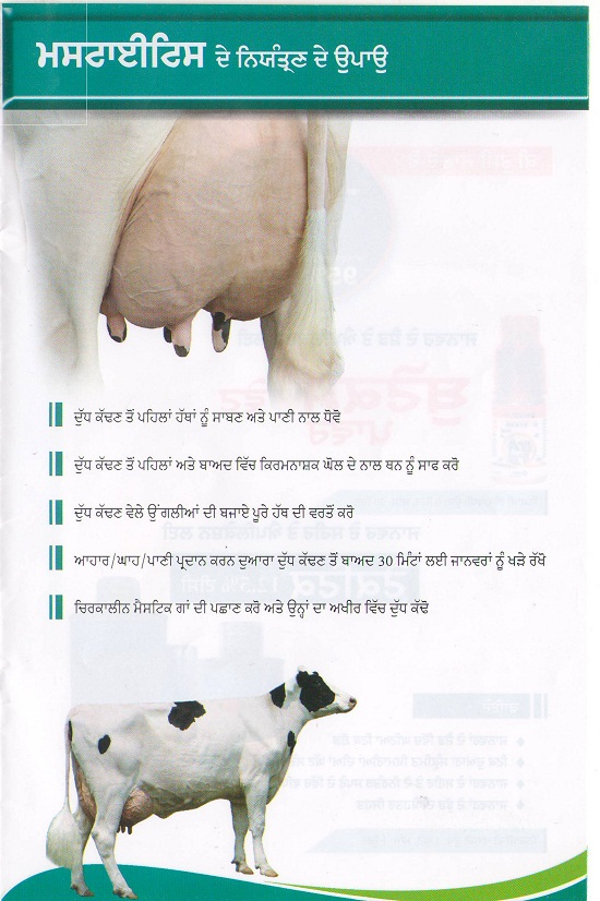
Intramammary infections are often described as subclinical or clinical mastitis. Clinical mastitis often involves hardening, reddening and swelling of the affected quarter. Milk secretion is often abnormal containing pus, fibrin (clots), and occasionally blood. The milk may also carry a pungent odour. The animal may be systemically ill. Subclinical mastitis involves infection without apparent signs of disease. Although transient episodes of abnormal milk or udder inflammation may appear, these infections are for the most part asymptomatic and, if the infection persists for months are termed chronic. Occasionally these infections persist for entire lactations or the lifetime of the cow.
Diagnosis
Detection is best done by examination of milk for somatic cell counts (predominantly neutrophils) using either the California Mastitis Test or automated methods provided by some dairies. Somatic cell counts are positively correlated with the presence of infection. Although variable (especially if determined on a single analysis), cows with a somatic cell count of ≥ 200,000 cells/mL have a high chance of being infected. Likewise, the higher the somatic cell count in a herd bulk tank, the higher the prevalence of infection in the herd. Causative agents may be identified by bacterial culture or PCR analysis of milk.
For information on control click here.
Control
Control of BVD is based on four equally important aspects:
- Selective culling – Reduction of circulating virus can be achieved with the introduction of a vaccination program and progressive culling of those animals that are identified as a potential source of the virus. In farms with a very low sero-prevalence (proportion that are positive on an antibody test) culling without vaccination can be an option. However in most farms due to the high sero-prevalence it is not economically feasible to test and cull all the sero-positive animals.
- Biosecurity – Maintaining biosecurity involves avoiding introduction of infected animals into the herd and/or implementing stict isolation / quarantine of introductions until proven negative, and restricting access of livestock to external sources of infection e.g. double fencing is in place at all perimeters, considering carefully sources of biological materials such as embryos, semen etc.
- Vaccination – The use of live vaccines is preferred above the inactivated ones because of the superior efficacy in clinical protection and more importantly in reduction of the virus circulation. For more on vaccination click here.
- Monitoring – This varies depending on the nature and risk status of your herd. Appropriate screening programmes can be discussed with your local veterinary practitioner.
Further information on the National control programme and IBR is available on the Animal Health Ireland website.
Vaccination
IBR marker vaccines are vaccines containing a gE deleted BoHV-1 strain for the vaccination of cattle against BoHV-1. Live and inactivated vaccines are available.
Recommendations for the choice between live and inactivated vaccine
The live vaccine has advantages in the following situations:
- Primary course
- Young animals
- High risk of infection
- Large herds or herds with regular introduction of animals
- Herds with high prevalence
- Intranasal route preferred when early onset of immunity is required
IBR Marker
Bovilis IBR marker live is an attenuated vaccine for active immunisation of cattle to reduce the intensity and duration of the clinical signs induced by an infection with BoHV-1 and to reduce excretion of field virus.
For vaccination schedule and data sheet details click here.
Bovilis IBR marker inac is an inactivated adjuvanted vaccine for active immunisation of cattle to reduce the intensity and duration of clinical respiratory signs induced by an infection with BoHV-1 as well as to reduce the nasal excretion of the field virus.
Comments are closed.